Treatments
Neuro Ophthalmology
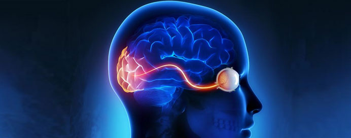
Where Eyes and Brain Connect
Your eyes are the windows to the world, but it’s your brain that interprets what you see. The optic nerves, eye muscles, and brain pathways work together in perfect harmony to create clear, meaningful vision. Neuro-Ophthalmology is a specialized field that deals with visual problems related to the nervous system.
Disorders affecting the optic nerves, eye movements, pupils, or brain areas controlling vision can lead to blurring, double vision, visual field defects, and even vision loss. Early detection and timely treatment are crucial, as many neuro-ophthalmic conditions may indicate serious underlying neurological or systemic diseases.
Common Neuro-Ophthalmic Disorders
Optic Neuritis
- Inflammation of the optic nerve, commonly linked to Multiple Sclerosis (MS).
- Symptoms: Sudden vision loss (usually in one eye), pain on eye movement, color vision changes.
- Requires urgent neurological evaluation.
Papilledema (Swelling of the Optic Disc)
- Caused by raised intracranial pressure (brain pressure).
- Symptoms: Headache, transient visual blackouts, double vision, nausea.
- Needs thorough evaluation to rule out brain tumors, idiopathic intracranial hypertension (IIH), or infections.
Optic Neuropathy (Ischemic / Compressive)
- Damage to the optic nerve due to reduced blood flow (Ischemic Optic Neuropathy) or compression by tumors, aneurysms.
- Sudden, painless vision loss or gradual deterioration.
- Requires urgent diagnosis to prevent permanent damage.
Oculomotor Nerve Palsies (3rd, 4th, 6th Cranial Nerve Palsies)
- Affect eye movements, causing double vision, droopy eyelid (ptosis), or abnormal head postures.
- Common causes: Diabetes, Hypertension, Trauma, Brain Aneurysms, Tumors.
- Some palsies resolve with observation; others may need surgical correction or prism glasses.
Myasthenia Gravis
- An autoimmune condition causing fatigable muscle weakness.
- Presents as drooping eyelids (ptosis), double vision (diplopia), or generalized weakness.
- Diagnosed with clinical tests, blood tests, and nerve conduction studies.
- Managed with medications and immunotherapy.
Visual Field Defects from Brain Lesions
- Stroke, tumors, aneurysms, or demyelinating diseases can affect brain areas responsible for vision.
- Symptoms: Loss of side vision (peripheral), top/bottom visual field, or quadrant vision loss.
- Requires brain imaging to find the underlying cause.
Pupillary Abnormalities (Anisocoria, RAPD)
- Unequal pupil sizes, sluggish light reactions may indicate nerve damage, optic nerve diseases, or brainstem disorders.
- Important clue to localize neurological issues.
Systemic Diseases & Neuro-Ophthalmic Problems
Certain systemic conditions can severely affect the optic nerves and eye-brain pathways:
- Diabetes & Hypertension: Nerve palsies, ischemic optic neuropathy.
- Thyroid Disorders (Thyroid Eye Disease): Eye movement restriction, double vision.
- Multiple Sclerosis (MS): Optic neuritis, visual pathway demyelination.
- Autoimmune Diseases (Lupus, Vasculitis): Inflammatory optic neuropathies.
- Infections (Tuberculosis, Syphilis, Viral): Optic nerve involvement.
Warning Signs You Should Never Ignore
- Sudden, painless loss of vision.
- Painful eye movements.
- Double vision or misalignment of eyes.
- Persistent headaches with visual disturbances.
- Drooping eyelid with/without double vision.
- Unequal pupil sizes.
- Transient blackouts or visual field loss.
Key Diagnostic Tests in Neuro-Ophthalmology
At Vision & Beyond, we offer advanced neuro-ophthalmic diagnostics including:
- Detailed Neuro-Ophthalmic Evaluation: Eye movements, pupil reactions, optic nerve head analysis.
- Visual Field Testing (Perimetry): Detects defects in peripheral and central vision.
- Optical Coherence Tomography (OCT) of Optic Nerve & Retinal Nerve Fiber Layer (RNFL): Non-invasive imaging for nerve damage assessment.
- MRI / CT Brain and Orbit Imaging: For suspected brain or orbital pathologies.
- Fundus Photography: To document optic disc changes.
- B-scan Ultrasonography: To assess optic nerve head swelling.
- Blood Tests & Autoimmune Panels: If systemic conditions are suspected.
Treatment Options for Neuro-Ophthalmic Disorders
- Corticosteroids & Immunotherapy: For optic neuritis, autoimmune optic neuropathies.
- Surgical Intervention: For compressive lesions, nerve decompression, or ptosis correction.
- Botox Injections: For nerve palsies causing eyelid spasms or facial dystonias.
- Prism Glasses or Strabismus Surgery: For persistent double vision.
- Management of Systemic Conditions: Diabetes, hypertension, infections.
- Neuro-Rehabilitation & Vision Therapy: For visual field loss and nerve palsies.
Early detection and accurate diagnosis play a life-saving role, as many neuro-ophthalmic symptoms can be the first sign of serious neurological diseases like stroke, brain tumors, or multiple sclerosis.
Frequently Asked Questions (FAQs)
-
What is a Neuro-Ophthalmologist?
A neuro-ophthalmologist is an eye specialist trained to diagnose and manage visual problems related to the nervous system, especially involving the optic nerves, brain pathways, and eye movement disorders.
-
How is double vision treated?
Treatment depends on the cause. It can range from prism glasses, Botox injections, strabismus surgery, or addressing the underlying neurological condition causing the problem.
-
What is optic neuritis, and can vision recover?
Optic neuritis is inflammation of the optic nerve, often linked to autoimmune diseases like MS. With prompt treatment (usually steroids), vision can recover, but some patients may have residual visual disturbances.
-
Can diabetes or hypertension cause nerve palsies?
Yes, microvascular cranial nerve palsies due to diabetes and hypertension are common and typically present with sudden onset double vision or droopy eyelid.
-
What is papilledema, and why is it dangerous?
Papilledema is swelling of the optic nerve head due to raised intracranial pressure. It is a serious sign that needs urgent evaluation to rule out brain tumors, infections, or cerebrospinal fluid (CSF) abnormalities.
-
Is it normal to have unequal pupil sizes?
A slight difference in pupil size can be normal, but if associated with other symptoms like droopy eyelid, headache, or vision changes, it may indicate a serious nerve or brain condition.
-
What is the role of MRI in neuro-ophthalmology?
MRI helps visualize the optic nerves, brain pathways, and surrounding structures, essential for diagnosing tumors, demyelinating diseases (like MS), or other neurological disorders affecting vision.
-
Can neuro-ophthalmic conditions cause permanent vision loss?
Yes. Delayed diagnosis and treatment can lead to irreversible optic nerve damage. However, many conditions, if detected early, can be managed effectively to preserve vision.
-
Can neuro-ophthalmic problems be prevented?
While some conditions like trauma-related nerve palsies or tumors are not preventable, maintaining control of diabetes, hypertension, and autoimmune diseases can significantly reduce the risk of neuro-ophthalmic complications.
Takeaway: Vision and Brain Work as a Team — Protect Both
Neuro-ophthalmic disorders often sit at the crossroads of eye and brain health. Recognizing the early warning signs and seeking specialized care can prevent permanent vision loss and may even detect serious neurological diseases early. At Vision & Beyond, we offer comprehensive neuro-ophthalmic evaluations, advanced imaging, and personalized treatment plans to safeguard your visual pathway.